
Figure 4. APS ameliorates doxorubicin-induced heart failure by restoring normal autophagy and decreasing apoptosis in vivo.
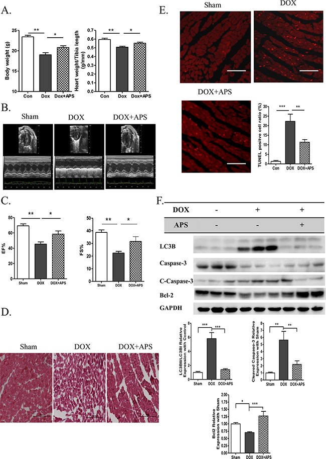
A. The average data of mouse body weight and heart weight/tibia length rate (g/mm) in sham, DOX, and DOX + APS groups 5 days after doxorubicin treatment (n = 8). B-C. Echocardiographic M-mode tracings and measurements of fractional shortening % (FS %) and ejection fraction % (EF %) 5 days after doxorubicin treatment (n = 8). D. Heart H&E staining of sham mice, doxorubicin-induced mice (DOX), and mice with APS pretreatment followed by doxorubicin treatment (DOX + APS) (n = 5, scale bar: 100 μM). E. TUNEL staining of apoptotic cells in the Sham, DOX and DOX + APS groups (n = 5, scale bar: 100 μM). F. Western blot and average data for LC3B II/I, Caspase-3, Bcl-2 in the sham, DOX, and DOX + APS groups (n = 5). (*P<0.05, **P<0.01, ***P<0.001).