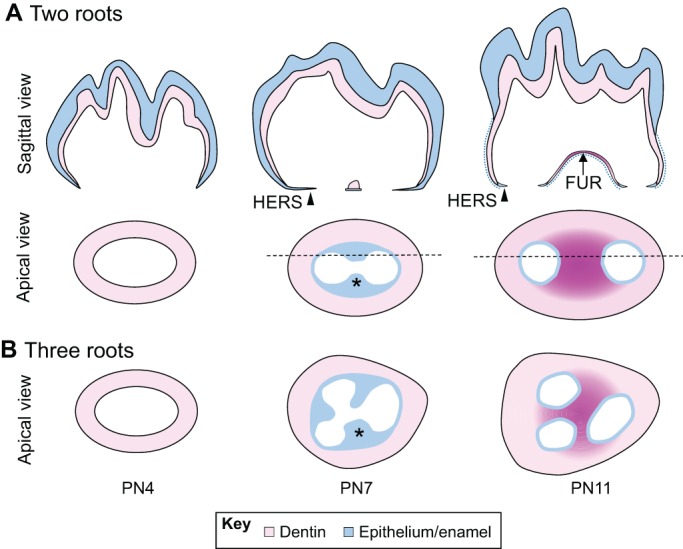
Fig. 3.

Development of the tooth root furcation in mice. (A) Schematics (sagittal view) of root furcation development in mice from PN4 to PN11. Epithelium-derived tissues, which include the ameloblast layer and enamel in the crown and the HERS in the root region, are depicted in blue. Dentin is depicted in pink. Arrowhead, HERS cells; blue dotted line, dissociated HERS cells. (B,C) Schematics (apical views) of root furcation development in two-rooted (B) and three-rooted (C) teeth in mice from PN4 to PN11. The HERS (blue) develops tongue-shaped epithelial protrusions (asterisks) that eventually fuse together to form the furcation (dark pink). Black horizontal dashed lines indicate the locations of the sagittal views shown in A.
