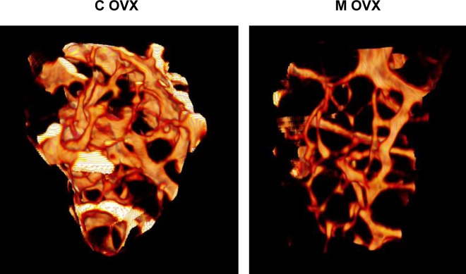
Figure 6. Illustration of 3D reconstructions of trabecular bone from ovariectomized control and mutant mouse using microCT.
3D reconstruction of distal femur trabecular bone was performed in eighteen month-old female Atg5Col1A-Cre+ (mutant, M) mice and their control littermates (control, C) following ovariectomy. These reconstructions based on 200 section analysis, illustrate the decrease of trabecular bone volume in mutant mice.

