
Figure 4. Effect of β-hydroxybutyrate on ER-stress-induced inflammasome formation via the AMPK pathway in vitro.
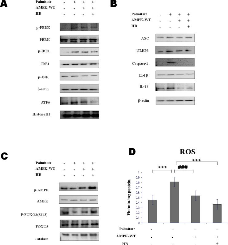
A. HepG2 cells were transfected with lentiviral particle wild-type AMPK. After 12 h, cells were treated with 1 mM β-hydroxybutyrate, followed by 250 μM palmitate for 1 h. Western blot analysis was performed to determine the levels of p-PERK, p-IRE1, p-JNK, and ATF6α. β-Actin and histone H1 blots are shown, to clarify the same amount of protein loaded in the cytosolic and nuclear fractions, respectively. B., C. HepG2 cells were transfected with lentiviral particle wild-type AMPK. After 12 h, cells were treated with 1 mM β-hydroxybutyrate, followed by 250 μM palmitate for 1 h. D. Reactive oxygen species generation was measured by dichlorofluorescein formation with the fluorescent probe 2′,7′-dichlorofluorescein diacetate. Results of one-factor ANOVA: ***p < 0.001 vs. control; ###p < 0.001 vs. palmitate. HB, β-hydroxybutyrate.