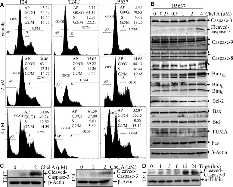
Figure 2. Chel A induces apoptosis in bladder cancer cell lines.
(A) After synchronization, the T24, T24T and U5637 cells were treated with Chel A as indicated for 24 hrs, and the cells were then fixed and subjected to cell cycle analysis by flow cytometry as described in “Materials and Methods”. The results represent one of three independent experiments. (B and C) After synchronization, the cells were treated with Chel A at the indicated concentrations for 24 hours or (D) Chel A at 4 μM for the time points indicated. The cell extracts were subjected to Western Blot with the indicated antibodies.

