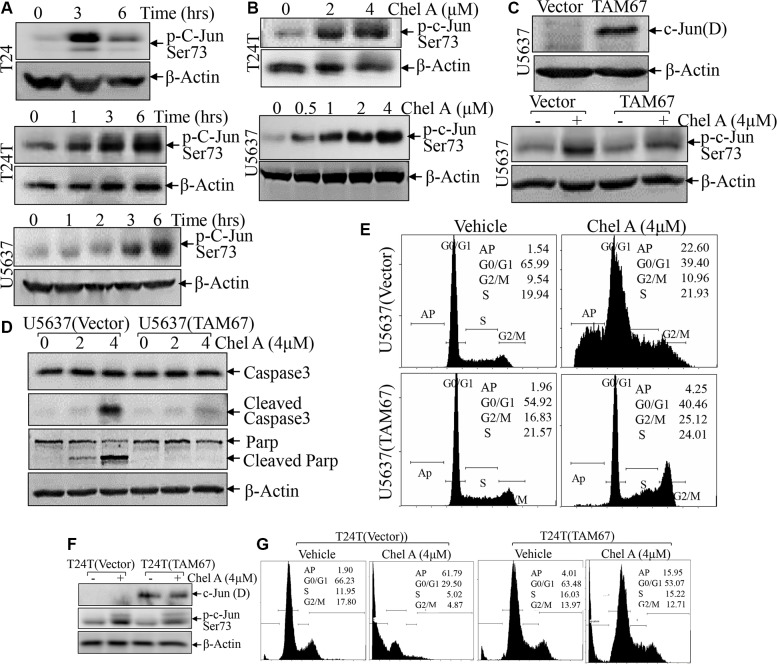
Figure 3. c-Jun phosphorylation up-regulated by Chel A plays a key role in the induction of bladder cancer cell apoptosis.
(A and B), After synchronization, T24 or T24T cells were treated with Chel A at 4 μM for at the time points indicated (A) or Chel A at the indicated concentrations for 6 hours (B). (C and F) T24T and U5637 cells stably transfected with TAM67 plasmids were seeded into 6-well plates. After synchronization, the cells were treated with or without 4 μM Chel A for 6 hrs. (D) U5637 cells stably transfected with TAM67 plasmids were seeded into each well of 6-well plates. After synchronization, the cells were treated with or without 4 μM Chel A for time indicated. The cell extracts were subjected to Western Blotting with the indicated antibodies. (E and G) The stable transfectants, T24(Vector) vs. T24T(TAM67) or U5637(Vector) vs. U5637(TAM67) cells, were treated with Chel A as indicated for 24 hrs, and the cells were then fixed and subjected to cell cycle analysis by flow cytometry. The results represent one of three independent experiments.

