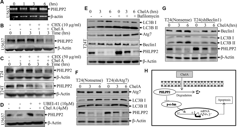
Figure 5. The down-regulation of PHLPP2 by Chel A is due to an increase in the rate of PHLPP2 protein degradation.
(A) Total RNA was isolated and subjected to RT-PCR analysis in the U5637 cells treated with 4 μM Chel A for the indicated time. PHLPP2 mRNA levels were evaluated by RT-PCR, with β-Actin mRNA levels used as loading control; (B) After synchronization, U5637 cells were pre-treated with MG132 for two hours. After MG132 was removed, cells were treated with or without 50 μg/ml CHX and/or 4 μM Chel A for time indicated, and the extracts were subjected to Western Blotting with anti-PHLPP2 or anti-β-Actin antibodies; (C) After synchronization, T24 or T24T cells were or were not exposed to 50 μg/ml CHX with or without 4 μM Chel A for the indicated time points, and the extracts were subjected to Western Blotting with anti-PHLPP2 or anti-β-Actin antibodies; (D) After synchronization, U5637 cells were respectively treated with or without 10 μM of UBEI-41 and with or without 4 μM of Chel A for three hours. The extracts were subjected to Western Blotting with anti-PHLPP2 or anti-β-Actin antibodies; (E) T24T cells were or were not incubated in 10 nM Bafilomycin A1 for 6 hours and treated with or without 4 μM Chel A. Cells were collected after time indicated and subjected to Western Blotting with anti-PHLPP2 or anti-β-Actin antibodies; (F) T24(shAtg7) and T24(Nonsense) were seeded into 6-well plates. After synchronization, the cells were treated with 4 μM Chel A for time indicated. The cell extracts were subjected to Western Blotting with the indicated antibodies. (G) T24(shBeclin1) and T24(Nonsense) were seeded into 6-well plates. After synchronization, the cells were treated with 4 μM Chel A for time indicated. The cell extracts were subjected to Western Blotting with the indicated antibodies. (H) The proposed model for the apoptotic responses following Chel A treatment.

