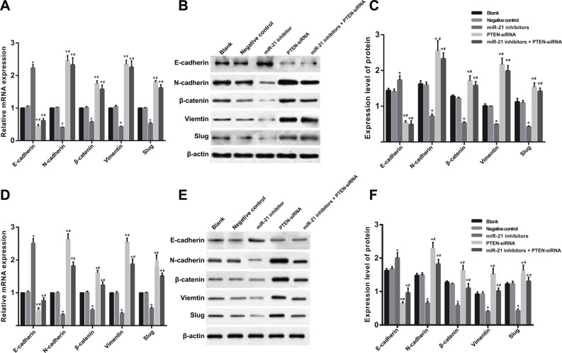
Figure 9. The mRNA and protein expressions level of EMT related factors in SGC-7901 and KATO-III cells after induced by TGF-β1 for 48 h (BL, blank group; NC, negative control group; IN, miR-21 inhibitors group.
(A) The analysis histogram of mRNA expression of EMT related factors in SGC-7901 cells; (B) The expression of EMT related factors in SGC-7901 cells tested by Western blot; (C) The analysis histogram of EMT related factors in SGC-7901 cells; (D) The analysis histogram of mRNA expression of EMT related factors in KATO-III cells; (E) the expression of EMT related factors in KATO-III cells tested by Western blot; (F) the analysis histogram of EMT related factors in KATO-III cells; TGF-β1, transforming growth factor β1; EMT, epithelial-mesenchymal transition; *comparison with the blank group and NC group, P < 0.05; #,comparison with IN group, P < 0.05).

