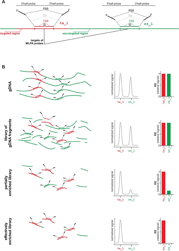
Figure 1. Strategy of MTTE analysis.

A. Schematic representation of targeted region-specific (ta_1; left-hand side) and non-targeted region-specific (nt_1; right-hand side) MLPA probes. Each MLPA probe is composed of two half-probes: a 5′half-probe and a 3′half-probe. Each half-probe is composed of a target-specific sequence (TSS), a primer-specific sequence (PSS), and a stuffer sequence (SS) that allows differentiating MLPA probes by size. More details about the design of MLPA probes may be found in [18, 20]. The first step of the MLPA reaction is hybridization of MLPA probes with the input DNA. Only probes which were correctly hybridized to their targets are subsequently ligated and then amplified with a pair of universal primers. The products of the MLPA reaction are separated in capillary electrophoresis and their relative signals are proportional to the dosage of their targets in the input DNA. B. The MTTE analysis of (from the top) gDNA, non-enriched gDNA library (reference), partially enriched library, and effectively enriched library. From the left, schematic representation of the MLPA probes hybridizing to targeted- and non-targeted regions in the input DNA (for simplicity, adapter sequences attached to DNA fragments during library preparation are not indicated in the scheme), electropherograms with signals (peaks) of ta_1 and nt_1 probes, bar-graphs showing relative signals of ta_1 and nt_1 probes.
