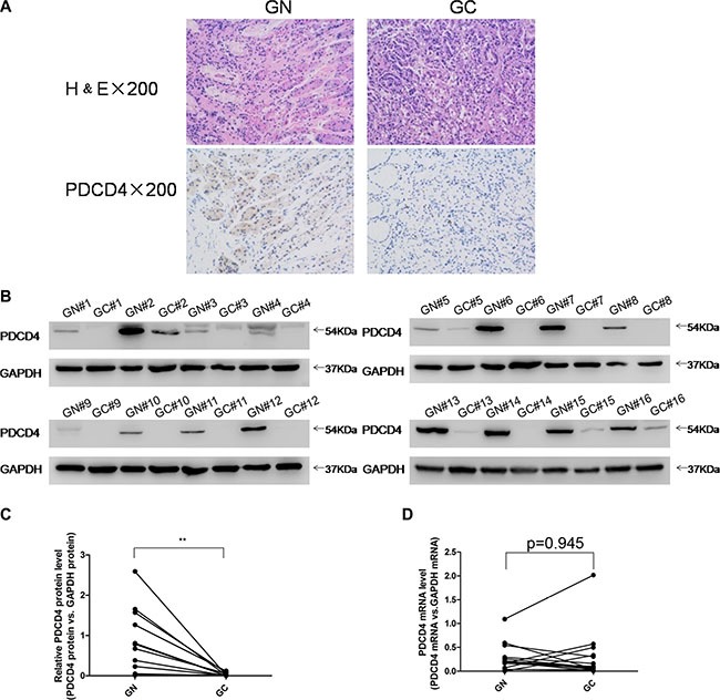
Figure 1. Expression patterns of PDCD4 in human gastric cancer tissues.

(A) H&E-stained sections and immunohistochemical staining for PDCD4 in gastric cancer tissue (GC) and gastric noncancerous tissue (GN) samples. (B and C) Western blotting analysis of PDCD4 protein levels in 16 pairs of GC and gastric GN samples. (A) representative image; (B) quantitative analysis. (D) Quantitative RT-PCR analysis of mRNA levels in 16 pairs of GC and GN samples (*P < 0.05; **P < 0.01).
