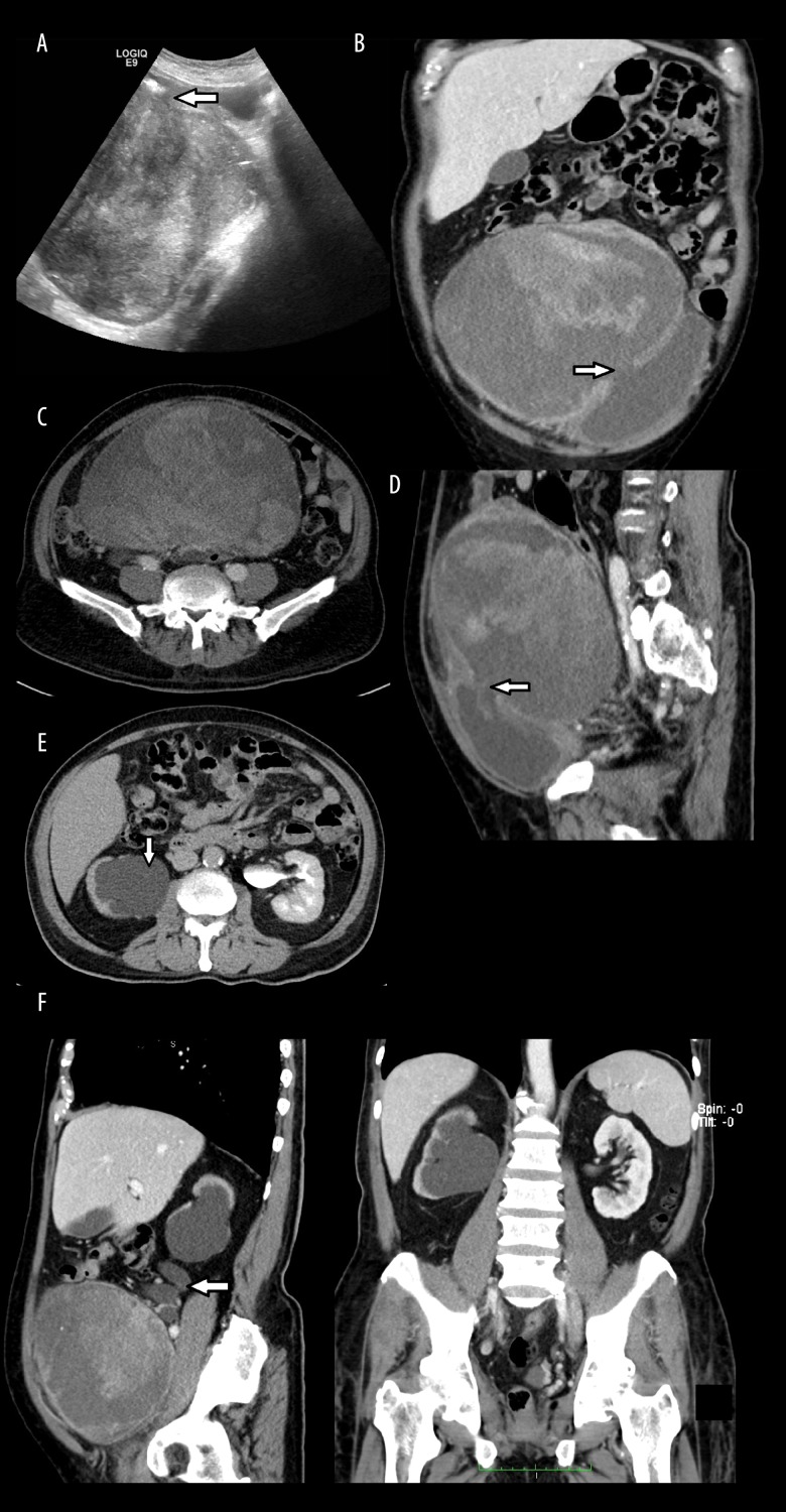
Figure 1.
Ultrasound and CT images. (A) Ultrasound images in sagittal view demonstrating the heterogeneous appearance of the intradiverticular bladder tumor. The neck of the diverticulum is noted (arrow). (B–D) Non-contrasted CT images on coronal (B), axial (C), and sagittal (D) views showing heterogeneity and huge size of the intradiverticular bladder tumor. The neck of the diverticulum is clearly shown on the coronal (B) and sagittal (D) views (arrow). (E) Contrasted CT image on axial view, during the excretory phase showing right hydronephrosis with absence of contrast excretion (arrow). (F) Contrasted CT images on sagittal and coronal views demonstrating right hydronephrosis. The right ureter is noted to be tortuous (arrow).

