
Figure 6. Inhibition of mitochondrial translation with Tigecycline provides a therapeutic window in a mouse model of Myc-induced lymphomas.
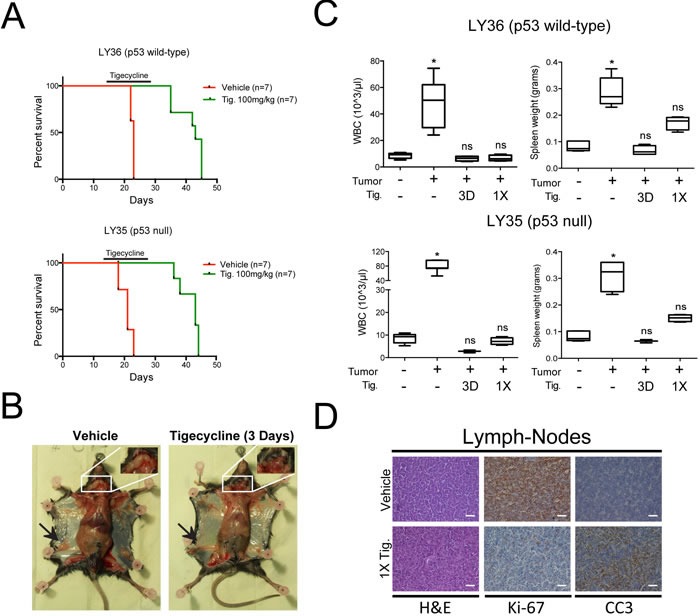
A. Kaplan-Meier survival curves of Eμ-myc lymphoma-transplanted mice treated with 100 mg/kg Tigecycline or vehicle (saline solution). 5*105 cells of either a p53 wt (LY36) or null (LY35) lymphoma were injected intravenously into C57Bl/6 recipient mice. Treatment with Tigecycline was started on day 14, when tumors were already palpable. Tigecycline was injected via intraperitoneal injection twice a day for 14 days. p < 0.001 for treated relative to untreated for both lymphomas, as determined by Log-rank (Mantel-Cox) test. B. Representative images of LY36 mice treated with vehicle or Tigecycline for three consecutive days. Note the absence of enlarged inguinal (black arrows) and cervical (white inset) lymph-nodes in Tigecycline-treated, as compared with vehicle-treated mice. C. Total white blood cell (WBC) count and spleen weight in vehicle and Tigecycline-treated mice after a single Tigecycline administration (1X) or 3 days of treatment (3D). Healthy C57Bl/6 mice were used as controls. Means, s.d. and statistical significance are as defined in Figure 2. D. Immunohistochemical stainings of lymph-nodes from mice treated with a single dose of tigecyline or vehicle. Representative H&E, Ki-67 and cleaved caspase 3 (CC3) images at 10X magnification are shown. Scale Bar 100 μm. See also Supplementary Figure S7.