
Figure 2. Codistribution of cytoplasmic actins with microtubules at the leading edge of MCF7 cells.
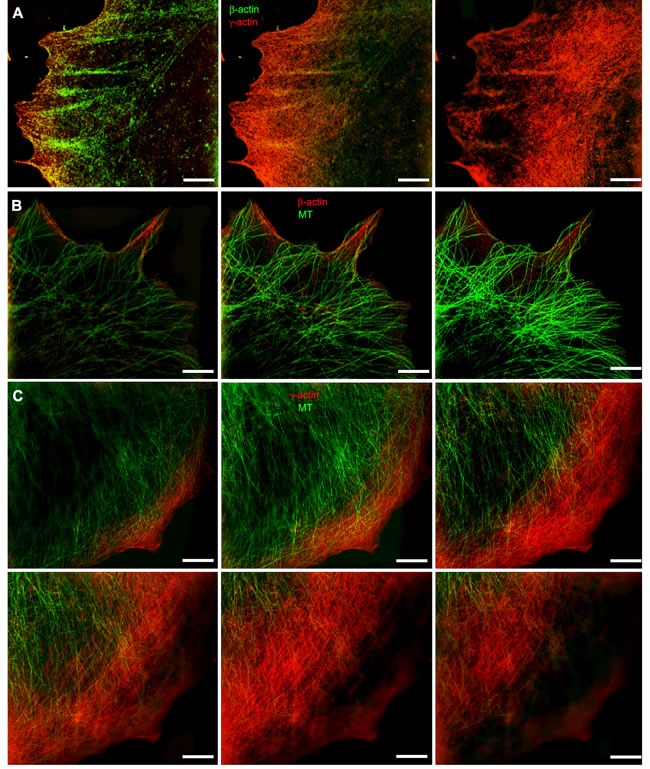
A. The β-actin bundles located at the basal level of the cell, closer to the substrate, and the cortical γ-actin network at the upper cell levels. B. Distribution of β-actin (basal level) and microtubules (upper level). C. Microtubules and γ-actin network codistribution. Microtubule tips are in close proximity to the γ-actin network. All panels represent galleries of optical sections taken with 0.12 μm step from the ventral (close to the substrate, first image) to the dorsal (last image) side of the lamella, SIM. Bars, 5 μm.