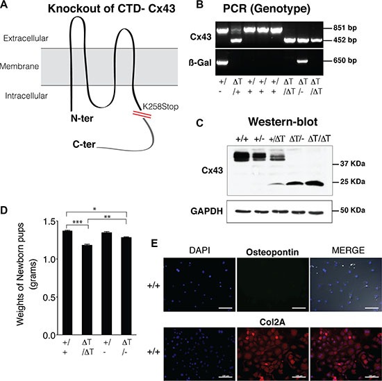
Figure 1. CTD-deficient cells display altered properties in their cell behaviour and phenotype.

(A) CTD-deficient mice (ΔT) express a carboxyl-terminal truncated Cx43 (K258Stop) instead of the WT Cx43 isoform [32]. (B and C) Mouse genotyping was performed by PCR and confirmed by western blot using an antibody raised against the N-terminal region of Cx43, in order to detect the full length (+/+ and +/−) and the truncated CTD mutants (2–4 day old mice). Cx43K258stop protein has a prolonged half-life time [32]. ß-Gal image represents the PCR amplicons for the ß-galactosidase that was inserted into the Cx43 allele to replace it. (D) CTD-mutant pups showing smaller body size than age and sex-matched wild type animals, were weighed as soon as they were born to avoid loss of water due to dehydration. Data is shown as mean ± S.E.M. (n = 9). (E) Osteopontin (marker of bone cells) and collagen type II (Col2A, chondrocyte specific marker) were determined by immunofluorescence on mouse primary knee articular chondrocytes isolated from wild-type mice. Scale bar represents 100 μm.
