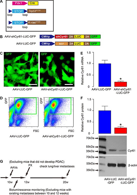
Figure 1. Schematic of in vivo experiment and AAV vectors.

(A) Schematic of triple mutant male mice (Pdx1-Cre, Conditional Loxp-STOP-Loxp (LSL)-Trp53R172H/+ and LSL-KrasG12D/+). (B) Schematic of AAV virus (AAV-shCyr61-LUC-GFP and AAV-LUC-GFP). (C) A human PDAC cell line, PANC-1, was transduced with AAVs and the cells appeared green due to expression of GFP in culture. (D) To obtain purified cells, the transfected cells were further subjected to flow cytometry to isolate GFP+ cells, shown by representative flow charts. (E–F) Cyr61 levels in the purified GFP+ cells by RT-qPCR for mRNA (E) and by Western blot for protein (F). (G) Mice received intraductal infusion of AAV at 10 weeks of age. The mice were kept for 2 weeks before pancreatectomy (PX) was performed. During the 2 weeks, bioluminescence was monitored and all mice that showed bioluminescence outside pancreas region (e.g. lung and liver) were excluded from the study. At the time of PX, mice that did not develop PDAC were also excluded from the study. Afterwards, the mice received insulin pellets and were kept for another 8 weeks, before they were lively analyzed for presence of distal metastases (at liver and lung) through bioluminescence and then sacrificed to analyze presence of distal metastases through examination of the GFP mRNA levels in liver and lung. *p < 0.05. N = 20. Scale bars are 10 μm.
