
Figure 4. Dependence of palmitate-induced tight junction disruption and enhanced permeability on caspase-1.
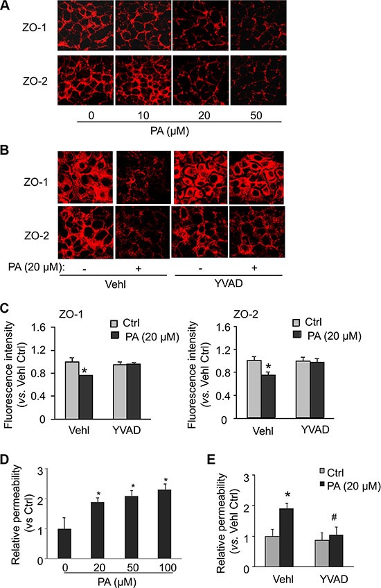
MVECs were stimulated with different concentrations of palmitate (PA: 0–50 μM) or with 20 μM palmitate in the presence of PBS (Vehl for vehicle) or caspase-1 inhibitor Ac-YVAD-CMK (YVAD: 10 g/ml) for 24 hours. (A and B) Representative fluorescence images show the cell membrane fluorescence of ZO-1 and ZO-2 from at least three independent experiments. (C) The protein expression of ZO-1 and ZO-2 were quantified by flow cytometry. Summarized data show the mean fluorescence intensity of ZO-1 or ZO-2 (n = 5–8). (D and E) MVECs on inserts of transwells were treated as above. Summarized data show the relative permeability of endothelial monolayers in the inserts for FITC-dextran (n = 4–5). *P < 0.05 versus Vehl Ctrl; #P < 0.05 vs. Vehl with PA.