
Figure 6. Inhibition of cathepsin B activity abolishes palmitate-induced Nlrp3 inflammasome activation, tight junction disruption, and enhanced permeability in MVECs.
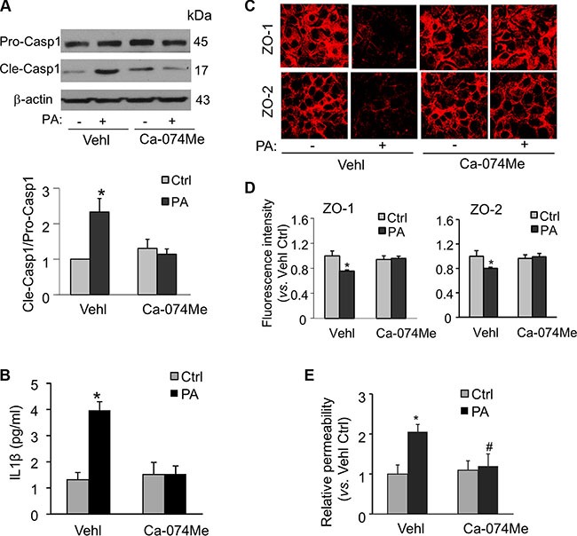
MVECs were stimulated with 20 μM palmitate in the presence of PBS (Vehl for vehicle) or cathepsin B inhibitor Ca-074Me (5 μM). (A) Western blot documents and summarized data showing the pro-caspase-1 (Pro-casp1) and cleaved caspase-1 (Cle-casp1) expression. (n = 4). (B) Summary of data for IL-1β production compared with untreated control (n = 5–8). (C) Representative fluorescence images show the cell membrane fluorescence of ZO-1 and ZO-2 from at least three independent experiments. (D) The protein expression of ZO-1 and ZO-2 were quantified by flow cytometry. Summarized data show the mean fluorescence intensity of ZO-1 or ZO-2 (n = 8). (E) Summarized data show the relative permeability of endothelial monolayers in the inserts for FITC-dextran (n = 3–4). *P < 0.05 versus Vehl Ctrl; #P < 0.05 vs. Vehl with PA.