
Figure 3. Anti-oxMIF mAbs sensitize cancer cells to cytotoxic drugs in vitro and in vivo.
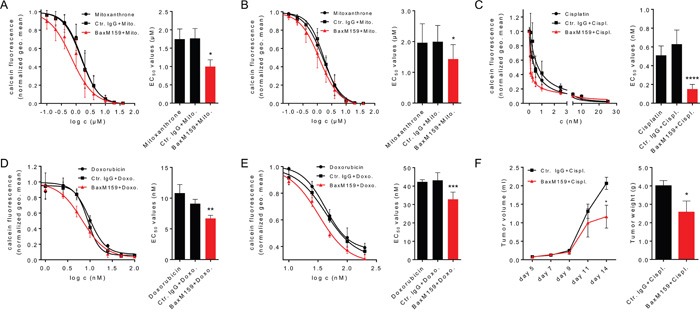
Prostate cancer cell lines LNCaP A. or PC3 B. were incubated with various concentrations of mitoxantrone (0.01-40 μM) either in the presence of 100 nM BaxM159, or matched human isotype control antibody (Ctr. IgG) or without antibody. The ovarian cancer cell line A2780 was incubated with various concentrations of cisplatin (0.1-25 nM) C. or doxorubicin (3.13-200 nM) D. either in the presence of 50 nM BaxM159, or matched human isotype control antibody (Ctr. IgG) or without antibody. The adriamycin-resistant ovarian cancer cell line A2780ADR E. was incubated with various concentrations of doxorubicin (3.13-200 nM) either in the presence of 50 nM BaxM159, or matched human isotype control antibody (Ctr. IgG) or without antibody. After 48 h cells were labeled with calcein-AM and live cells were counted by flow cytometry. EC50 values for cytotoxic drugs were calculated by fitting the data points to a four-parameter variable slope equation (Hill-equation). Curve fits (left panels) and EC50 values (right panels) are represented as means ± SEM from at least 4 independent experiments. *p<0.05; **p<0.01; ***p<0.001; ****p<0.0001. We used one-way ANOVA followed by Dunnett's multiple comparison test. F. MF-1 nude mice (n= 10/group) were inoculated with 1 × 10⁶ A2780 cells suspended in matrigel. Mice were treated with cisplatin (2.5 mg/kg) and BaxM159 (15 mg/kg) or human Ctr. IgG (15 mg/kg). Tumor volumes were measured at indicated time points. On day 14, mice were sacrificed, the tumors excised and weighed. Data are shown as means ± SEM. *p<0.05. We used student's unpaired t-test.