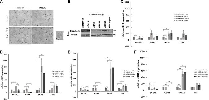
Figure 5. RNAi-mediated inhibition of BCL9L expression counteracts epithelial-mesenchymal transition in pancreatic cancer cells treated with TGF-β.

(A) Panc-1 cells stably transduced with control vector or BCL9L shRNA (shBCL9L) were treated with 5 ng/ml TGF-β or left untreated for 72 h and subsequently visualized using a phase-contrast microscope. Control cells responded to TGF-β treatment by adopting a mesenchymal, spindle-like phenotype whereas BCL9L-knockdown cells largely retained the cobblestone-like epithelial morphology. (B) Western Blot analysis of E-cadherin and α-tubulin (loading control) protein levels in Panc-1 cells treated with 5 ng/ml TGF-β for 72 h. shRNA- and siRNA-mediated knockdown of BCL9L (shBCL9L, siRNA1, siRNA2, siRNA pool) induced an upregulation of E-cadherin expression in comparison to controls (Vector ctrl, siCTR). Shown are representative images of 2 experiments. mRNA expression levels of epithelial (CDH1) and mesenchymal (SNAI2, VIM) genes were quantified in BCL9L-knockdown and control Panc-1 cells treated or not with TGF-β for 24 h (C), 48 h (D), 72 h (E) and 96 h (F), respectively. Data shown represent mean and standard deviation of 3 independent experiments. *p ≤ 0.05, **p ≤ 0.01, ***p ≤ 0.001 (Student's t-test).
