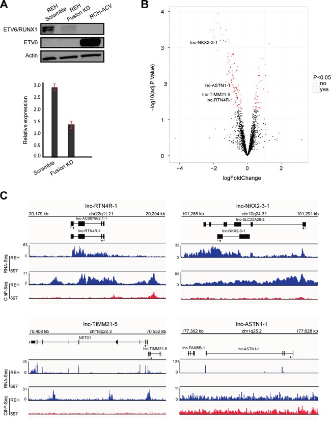
Figure 2. Identification of ETV6/RUNX1 regulated lncRNA.

A. Western blot (top) and RT-qPCR (below) analysis of ETV6/RUNX1 protein and transcript in REH cells upon fusion knockdown (KD). RCH-ACV used as an ETV6/RUNX1-negative B-ALL cell line with wild type ETV6. This result is representative for three independent biological replicates. B. Volcano plot representing the differentially expressed lncRNAs (red) when comparing fusion knockdown to the scramble (adjusted p-value < 0.05). C. RNA sequencing tracks (RNA-Seq) and H3K27ac binding patterns (ChIP-Seq) for lnc-NKX2-3-1, lnc-TIMM21-5, lnc-ASTN1-1 and lnc-RTN4R-1 in REH and 697 cell lines.
