
Figure 1. FGF4, not FGF7 treatment induces EMT, causes a switch from FGFR2 IIIb to FGFR2 IIIc and changes the cell morphology and behavior of A549 and H1299 lung ADC cells.
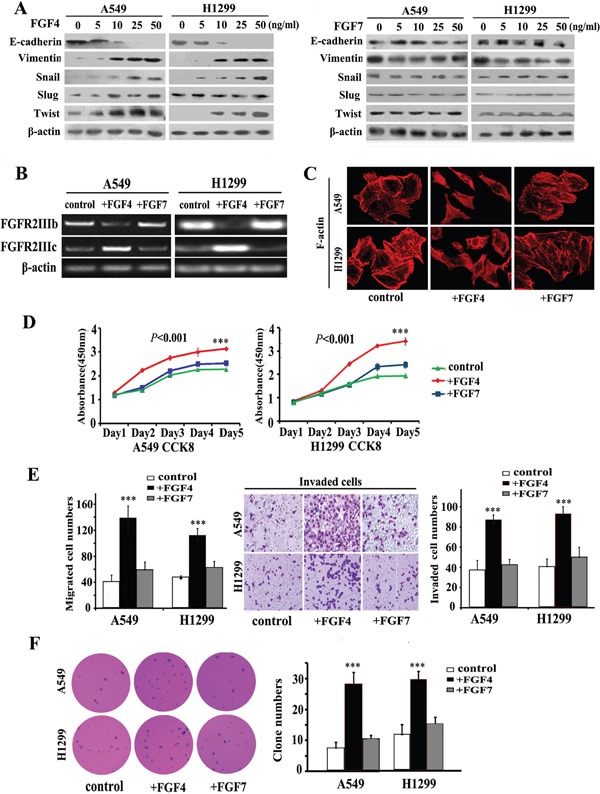
A. Western blot assay measuring the expression of epithelial protein (E-cadherin), mesenchymal protein (Vimentin), and EMT transcription factors (Snail, Slug, and Twist) in A549 and H1299 cells treated with FGF4 and FGF7 at different concentrations of 0, 5, 10, and 25, and 50 ng/mL, respectively. B. RT-PCR was performed to detect gene expression alterations in FGFR2 IIIb and FGFR2 IIIc. C. Cell morphology was analyzed by confocal laser scanning microscopy according to the immunolocalization of F-actin. D. CCK-8 cell proliferation assay was conducted in cells upon incubation with FGF4 or FGF7 in 1, 2, 3, 4, and 5 d, respectively. E. Transwell assay showing migration/invasion of cells treated with FGF4 or FGF7. F. Anchorage-independent growth was conducted to show the colony-initiation ability of cells exposed to FGF4 or FGF7. A549 and H1299 cells were treated with FGF4 and FGF7 at 10 ng/mL for 24 h, respectively. The control is A549 and H1299 cells without FGF4 or FGF7 stimulation. All graphs represent the mean ± SD of three independent experiments. The axis represents the fold change in the number of cells. ***P < 0.001.