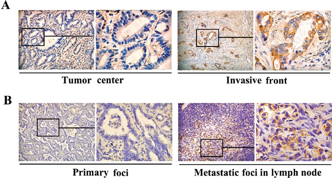
Figure 6. Differences in FGF4 levels within human ADC tissues and in matched lung ADC primary foci and lymph node metastatic foci.

Immunohistochemical staining of FGF4 showed A. different FGF4 levels in the tumor center and the invasive front of the same lung ADC tissue sample (200×), B. different FGF4 levels inlung ADC primary foci and lymph node metastatic foci from a same patient (200×).
