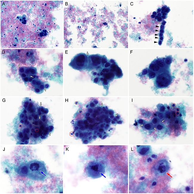
Figure 1. Liquid-based cytological findings of cervical glassy cell carcinoma.

A. Thinprep preparation of the cervical sample shows amphophilic, granular necrotic debris (tumor diathesis) and a few small clusters of polygonal tumor cells. B. The tumor cell clusters or individually scattered tumor cells are unevenly distributed. Tumor diathesis is apparent. C. Although some tumor cells display endocervical-like pseudocolumnar arrangements (black arrowheads), there is no definite evidence of glandular differentiation. A white arrow indicates the intercellular window. D. The tumor cells have relatively fine chromatin and prominent, solitary nucleoli. Abundant, cyanophilic cytoplasm and discrete cell borders (white arrows) are evident. E. Under high-power magnification (×400), the tumor cells show large, oval to round, pleomorphic nuclei and “intercellular windows” produced by discrete cytoplasmic outlines and cytoplasmic molding (white arrow). There are no intercellular bridges. F. Tumor cells are 3–7 fold larger than lymphocytes or neutrophils. Chromatin distribution irregularities, hyperchromasia, and significant anisonucleosis are apparent. G-I. In several areas, an intimate admixture of neutrophils (red circles) and tumor cells, so-called granuloepithelial complexes, is seen. Cytoplasmic molding and intercellular windows (white arrows) are observed. J-K. Mitotic figures (blue arrows) are present. L. Atypical mitotic figures (red arrow) are also detected (A-L, Papanicolaou stain).
