
Figure 2. Verification of recombinant VACVs structure.
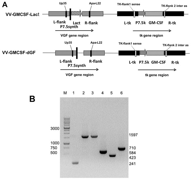
A. – schematic view of VV-GMCSF-Lact and VV-GMCSF-dGF (control variant) recombinant virus genomes with primer positions indicated. B. – PCR identification of recombinant VACVs DNA with primers TK-flank1 sense and TK-flank 2 inter as (Lanes 1–3) and with primers Up35 and Apa-L22 (Lanes 4–6). Lanes 1 and 4 are wild-type VACV (L-IVP); 2 and 5 – VV-GMCSF-dGF; 3 and 6 – VV-GMCSF-Lact. M – DNA molecular weight marker.