
Figure 3. Expression of virus mediated transgene proteins in infected CV-1 cells.
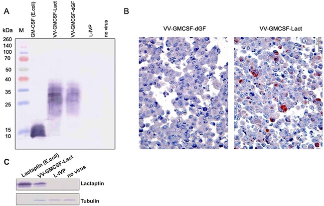
A. – Western blot analysis of human GM-CSF in cell culture medium. M – protein molecular weight marker. CV-1 cells were infected with VV-GMCSF-Lact, VV-GMCSF-dGF or wild type L-IVP (negative control). Recombinant GM-CSF expressed in E.coli was used as a positive control. Medium from non-treated cells was also used as a negative control. B. – immunohistochemical staining of CV-1 cells infected with recombinant VACVs. Representative paraffin sections of CV-1 cells were treated with anti-lactaptin antibodies and AEC chromogen (red colored cells), counterstained with hematoxylin. C. – Western blot analysis of lactaptin expression in infected cells. CV-1 cells were infected with VV-GMCSF-Lact or wild type L-IVP (negative control). Recombinant lactaptin expressed in E.coli was used as a positive control. One representative of the two independent experiments is shown.