
Figure 5. The influence of recombinant VACVs on cell proliferation.
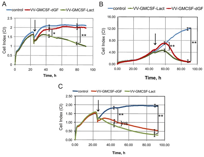
iCELLigence data showing typical Cell Index curves (CI) that reflect cell proliferation in real-time mode. Cells were seeded at 1500 cells per well and 24 or 46 h later recombinant VACVs were added to the wells. The point at which VACVs were added is indicated by the arrow. A, B and C. - 0.1, 0.5 and 10 PFU/cell, respectively. One representative of two independent experiments is shown. The difference between groups was statistically significant at *p<0.05 and **p<0.01 and non significant at p>0.05 (ns).