
Figure 6. Features of apoptosis of the MDA-MB-231 cells after recombinant VACVs infection.
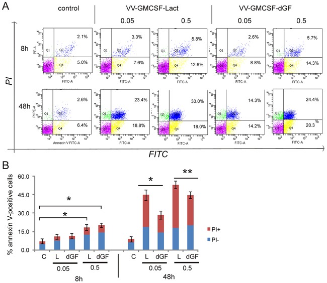
MDA-MB-231 cells were treated with recombinant VACVs (0.05 and 0.5 PFU/cell) or with saline (control) for 8 and 48 h and then cells were stained using annexin V/propidium iodide (PI). The stained cells were assayed for apoptosis by flow cytometry. Cell populations with the annexin V−/PI− phenotype (Q3) were designated as living cells, annexin V+/PI− (Q4) - as apoptotic cells, and annexin V+/PI+ (Q2)- as secondary necrotic cells. A. – One representative of three independent experiments is shown. B. – Bar graph summarized the percentage of apoptotic cells from three independent experiments (*p<0.01, **p<0.05). C-control, dGF- VV-GMCSF-dGF, L - VV-GMCSF-Lact.