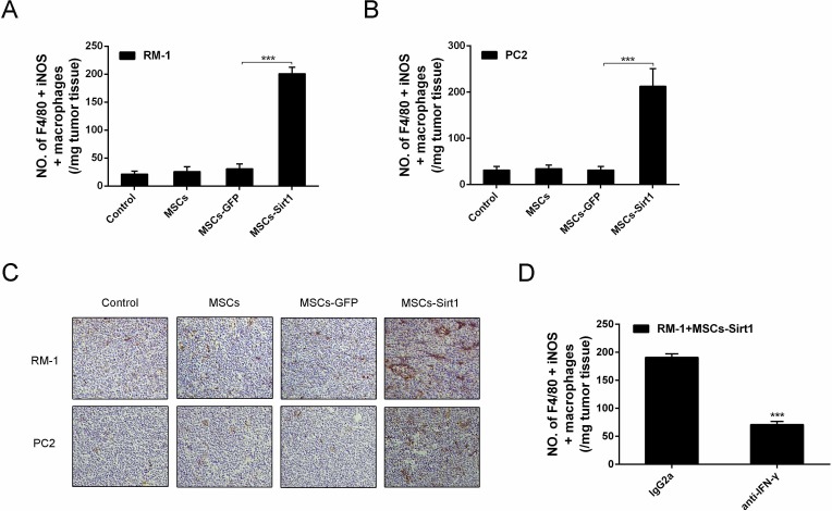
Figure 6. Macrophages activation in tumor regions.
Tumor infiltrating cells were isolated from A. RM-1 and B. PC2 tumors, then stained with anti-F4/80 antibody (Abcam), followed by intracelluar staining with anti-iNOS antibody. C. Immunohistochemistry against CD68 and iNOS in tumour tissues was observed by microscope (original magnification: ×200). D. Mice were intraperitoneally injected with anti-IFN-γ mAb or control lgG2a per 2 days until tumors excision. Tumor infiltrating cells were isolated and performed flow cytometric analysis. Each group consists of 6 mice. ***, P < 0.001.

