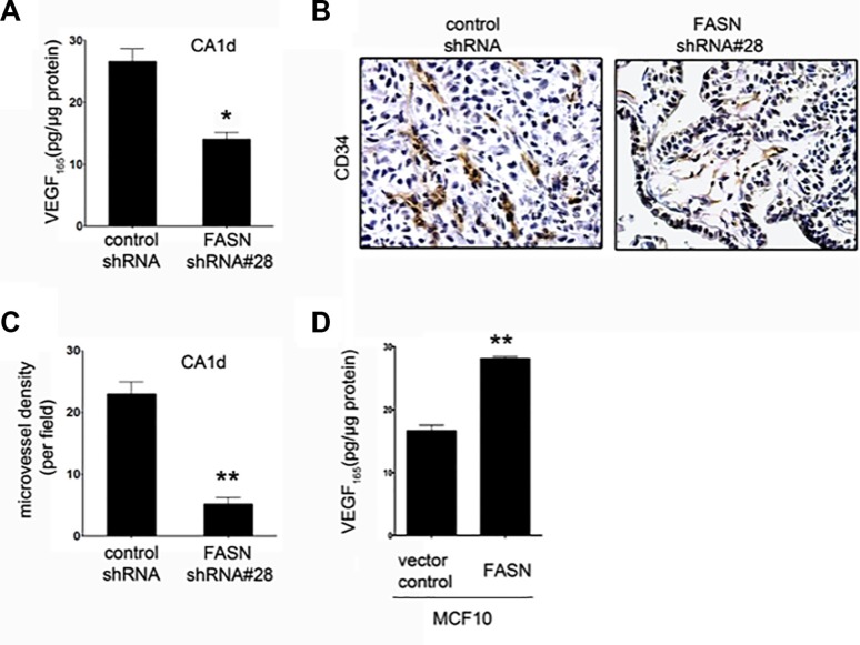
Figure 3. FASN depletion reduces VEGF secretion and angiogenesis in CA1d cells.
(A) ELISA-based analysis of VEGF in the supernatant from CA1d/vector control or FASN-deficient CA1d (CA1d/FASN shRNA#28) cells. Note that FASN knockdown results in a significant decrease in secreted VEGF. Data from one representative experiment are presented as mean ± SD; *p = 0.02. (B) Visualization of vascularization in tumors from control (vector) and FASN-depleted (FASN shRNA#28) xenografts by immunohistochemical staining for CD34. Note the reduced vasculature in FASN-deficient tumors compared with control tumors. (C) Quantification of one representative experiment as described in B. Results are presented as mean ± SD; **p < 0.001. (D) FASN overexpression increases VEGF secretion in non-transformed MCF10 cells. ELISA of VEGF in supernatant from MCF10 vector control or FASN over-expressing cells. Data from one representative experiment are presented as mean ± SD; **p < 0.001.

