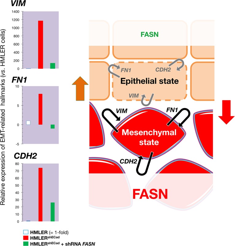
Figure 5. Transient knockdown of FASN gene expression suppresses structural hallmarks of EMT.
Left. Total RNA from HMLER, HMLERshECad, and HMLERshEcCad + shRNA FASN cells was characterized in technical triplicates for the relative abundance of 19 mRNAs whose levels, in published results [76, 77], are significantly altered during activation/deactivation of the EMT genetic program. The transcript abundance of selected EMT-related genes (VIM, FN1, and CDH2) was calculated using the delta Ct method and presented as fold-change vs. basal expression in HMLER cells. Right. When breast epithelial cells acquire a fibroblast-like morphology during EMT, they not only down-regulate the obvious effectors of the epithelial phenotype such as E-cadherin (CDH1), but also switch the expression of an entire battery of genes including those that encode mesenchymal intermediate filaments (e.g., cytokeratins à vimentin switch) and ECM proteins (e.g, fibronectin, FN1). The status of FASN signaling appears to coincide with a self-reinforcing attractor (see Figure 7) in the structural configuration of the breast epithelial cell cytoskeleton, thus explaining that the sole correction of exacerbated lipogenesis can stably reprogram the malignant, invasive phenotype of cancer cells back to normal-like tissue architectures.

