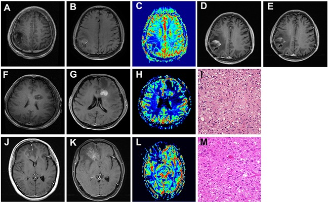
Figure 4. The clinical application of CTC in distinguishing tumor recurrence from radionecrosis.

A, F, G. Contrast axial T1-weighted image. After gross total resection, there is a surgical cavity without enhancement. B, G, K. Contrast axial T1-weighted image. After completion of RT, there is a new enhancing mass lesion on the initial post-RT MRI. C, H, L. rCBV map showed hypoperfusion (C and L, Patient 1 and Patient3) or hyperperfusion (H in patient 2) of the enhancing lesion (ROI 1) when compared to the contralateral normal white matter (ROI 2). D. and E. Follow-up of MRI performed 3(D) and 4(E) months after the initial post-RT MRI (Patient 1). I, M. Pathological findings in the second operation: tumor recurrence of GBM (Patient 2 and 3, HE, 10 x10). Patient 1: image A-E. Patient 2: image F-I. Patient 3: image J-M.
