
Figure 3. Cytokeratin and vimentin expression in LLC cells.
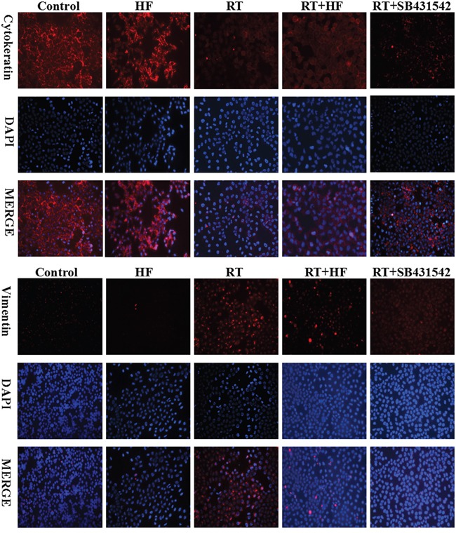
Immunofluorescence analysis of cytokeratin (red) and vimentin (red) expression in LLC cells, counterstained with DAPI (blue). Cells were stained 48 hours after indicated treatments.
Official websites use .gov
A
.gov website belongs to an official
government organization in the United States.
Secure .gov websites use HTTPS
A lock (
) or https:// means you've safely
connected to the .gov website. Share sensitive
information only on official, secure websites.
Immunofluorescence analysis of cytokeratin (red) and vimentin (red) expression in LLC cells, counterstained with DAPI (blue). Cells were stained 48 hours after indicated treatments.