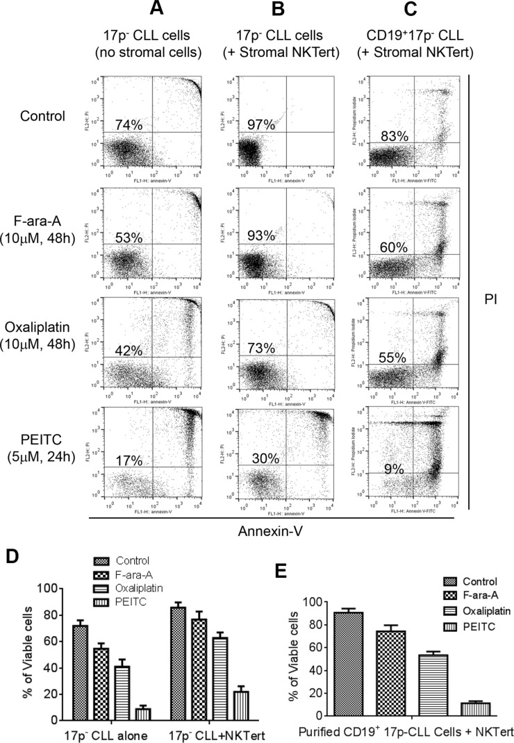
Figure 1. Comparison of cytotoxic effect of PEITC and standard chemotherapeutic agents in primary CLL cells with 17p deletion.
(A) Cell death induced by F-ara-A (10 μM, 48 h), Oxaliplatin (10 μM, 48 h), or PEITC (5 μM, 24 h) in primary 17p- CLL cells cultured alone (without stromal cells). Cell viability was analyzed by flow cytometry after double staining with Annexin V-PI. Representative dot plots of independent experiments using 9 different CLL patient samples are showed (n = 9). (B) Cell death induced by F-ara-A (10 μM, 48 h), Oxaliplatin (10 μM, 48 h), or PEITC (5 μM, 24 h) in 17p- CLL cells co-cultured with human bone marrow stromal NKTert cells. Cell viability was analyzed by flow cytometry after double staining with Annexin V-PI. Representative dot plots of independent experiments using 9 different CLL patient samples are showed (n = 9). (C) Cell death induced by F-ara-A (10 μM, 48 h), Oxaliplatin (10 μM, 48 h), or PEITC (5 μM, 24 h) in purified 17p- CD19+ CLL cells co-cultured with human bone marrow stromal NKTert cells. Cell viability was analyzed by flow cytometry after double staining with Annexin-V/PI. Representative dot plots of 3 independent experiments using 3 different CLL patient samples are showed (n = 3). (D) Quantitative comparison of cell death induced by F-ara-A (10 μM, 48 h), Oxaliplatin (10 μM, 48 h), or PEITC (5 μM, 24 h) in 17p- CLL cells alone or co-cultured with NKTert cells. (E) Quantitative comparison of cell death induced by F-ara-A (10 μM, 48 h), Oxaliplatin (10 μM, 48 h), or PEITC (5 μM, 24 h) in purified 17p- CD19+ CLL cells co-cultured with NKTert cells.

