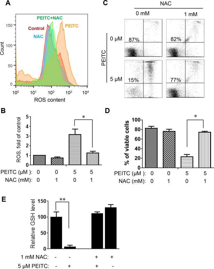
Figure 4. Effect of NAC on PEITC-induced ROS accumulation and cell death in primary CLL cells with 17p-deletion.
(A) Effect of NAC on PEITC-induced ROS accumulation in primary 17p- CLL cells. 17p- CLL cells were treated with 1 mM NAC for 1 hour before exposure to 5 μM PEITC for 2 h. Cellular ROS was analyzed by flow cytometry after staining with DCF-DA. Data shown are representative of 6 independent experiments. (B) Bar graphs showing quantitative analysis of ROS in CLL cells treated with PEITC, NAC, and their combination. *p < 0.05 (n = 6). (C) Effect of NAC on PEITC-induced cell death in primary CLL cells 17p- CLL cells with 17p-deletion were treated with 1 mM NAC for 1 hour before exposure to 5 μM PEITC for 24 h. Cell viability was analyzed by flow cytometry after double staining with Annexin V-PI. (D) Bar graphs showing quantitative analysis of cell death in primary CLL cells treated with PEITC, NAC, and their combination. *p < 0.05 (n = 6). (E) Depletion of cellular glutathione by PEITC treatment (5 μM, 6 h) in primary CLL cells with 17p-deletion in the presence or absence of 1 mM NAC. **p < 0.01 (n = 6).

