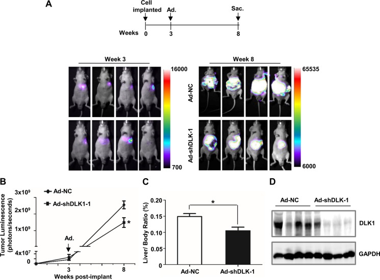
Figure 3. DLK1 knockdown mediated by adenovirus administration inhibits orthotopic xenograft tumor growth in athymic mice.
(A) Scheme and representative pseudocolor images of orthotopic xenograft tumors within nude mice at 3 and 8 weeks after inoculation. 2 × 10⁶ Huh-7 cells expressing luciferase were injected into the left liver lobe of nude mice. Luciferase imaging of these mice was performed once a week until tumor luminescence was observed, and then recombinant adenoviruses were administered at week 3 post cell implantation by tail vein injection. Luminescence imaging of these mice was performed at week 8 after inoculation. (B) Tumor luminescence was analyzed. Data represent the mean of 4 mice for each group ± sem (error bars). (C) Liver/body weight ratio was determined when these nude mice were sacrificed. (D) Western blotting assay was used for evaluating the efficiency of adenovirus-mediated DLK1 knockdown in these orthotopic xenograft tumors. Adenoviral vector containing scrambled shRNA was used as negative control. *p < 0.05.

