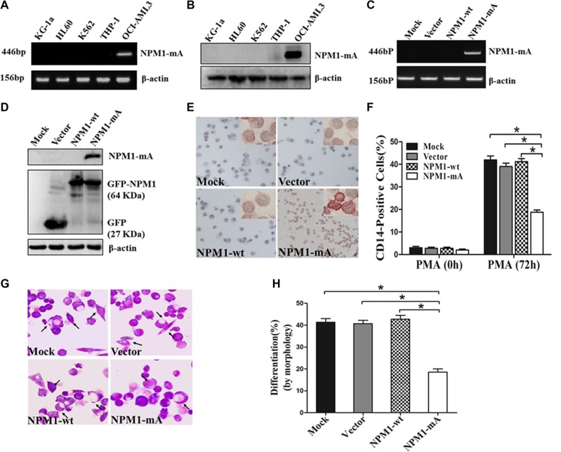
Figure 1. Overexpression of NPM1-mA inhibits myeloid differentiation of THP-1 cells.
(A–B) RT-PCR and western blot showing the absence of NPM1-mA mRNA and protein expression in KG-1a, HL60, K562 and THP-1 cells. OCI-AML3 cells were included as positive control. β-actin served as the loading controls. (C–D) NPM1-mA expression in THP-1 cells transfected with the recombinant plasmids expressing NPM1-mA, NPM1-wt or empty vector, measured by RT-PCR and western blot. (E) Representative results of cytoplasm-dislocated NPM1 mutant protein detected by immunocytochemistry staining (APAAP) in NPMc+ cells of the NPM1-mA group (×200). (F) The expression of CD14 in transfected THP-1 cells followed by PMA induction for 72 h was determined by flow cytometry. (G) Representative Wright-Giemsa staining images of transfected cells induced by PMA for 72 h. The arrows point to differentiated cells. (H) Percentage of differentiated cells determined by counting at least 200 cells on the slides under a light microscope. Three independent experiments were performed. *P < 0.05.

