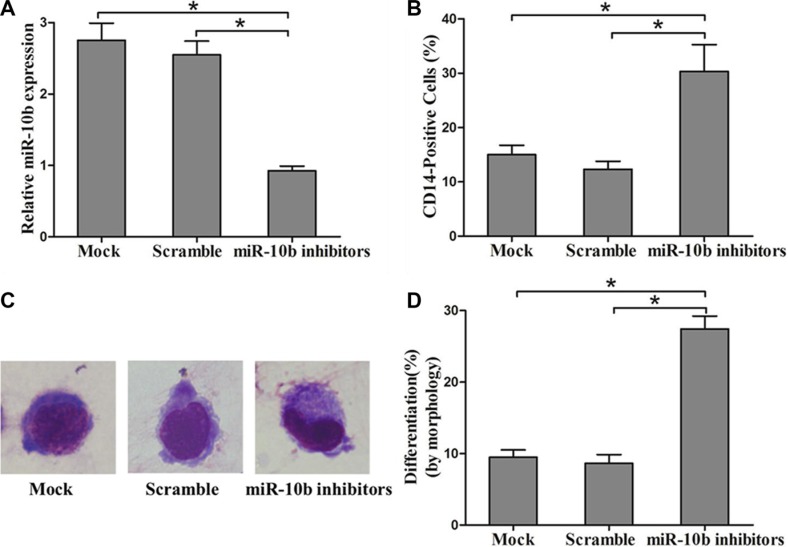
Figure 5. Suppression of miR-10b promotes myeloid differentiation of OCI-AML3 cells.
(A) Levels of miR-10b in OCI-AML3 cells transfected with miR-10b scramble or inhibitors measured by qRT-PCR. (B) Expression of CD14 in transfected OCI-AML3 cells under PMA treatment for 72 h as determined by flow cytometry. (C) Representative Wright-Giemsa staining image of transfected OCI-AML3 cells under PMA treatment for 72 h. (D) Percentage of differentiated cells determined by counting at least 200 cells under a light microscope. Three independent experiments were performed, *P < 0.05.

