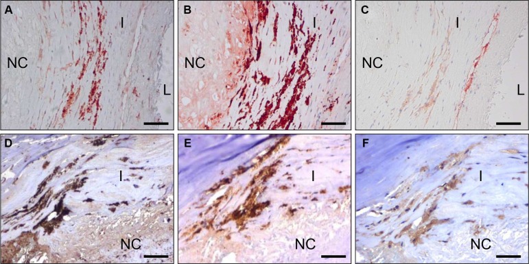
Figure 4. C-C7 recognizes blood vessels and macrophages in human atherosclerosis.
Immunostainings of surgical samples of atherosclerotic lesions from carotid arteries with CC7 (A, D) the macrophage marker CD68 (B, E) and the endothelial marker CD31 (C) and anti-dynactin-1 p150Glued (F, the anti-dynactin antibody was implemented in thee staining based on identification of dynactin as a binding partner of C-C7). (A–C) and (D–F) represent serial sections from two different lesions. Note the similarity in staining profiles for C-C7 and dynactin P150Glued (D and F, respectively). Tissues in A–C were stained with AEC, tissues in (D–F) with DAB. NC = nectrotic core, I = intima, L = lumen. Bars correspond to 100 μM in panels (A–C) and to 50 μm in panels (D–F).

