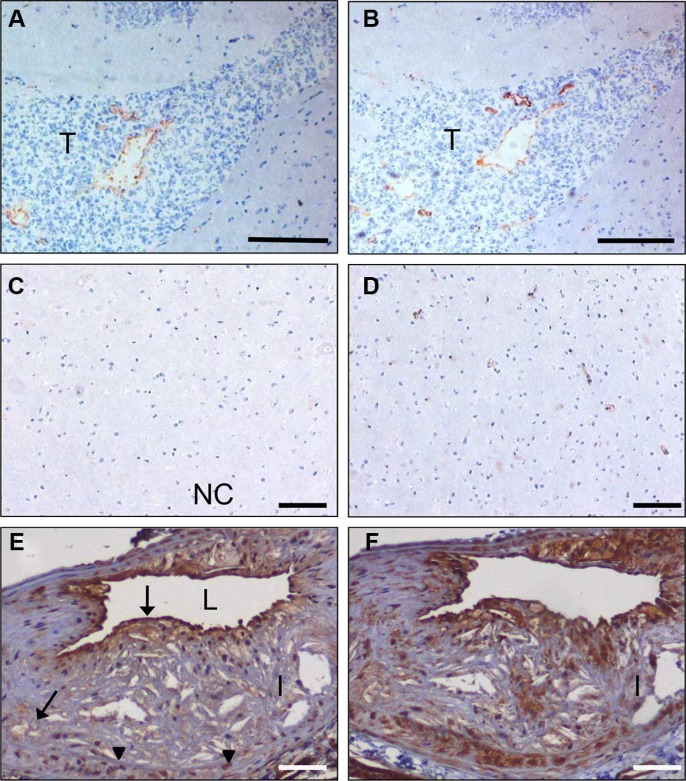
Figure 6. In vivo targeting of glioma xenografts and atherosclerotic lesions by C-C7-displaying M13 phages.
Monoclonal C-C7 phages were injected intravenously in mice carrying orthotopic E98 xenografts and phage distribution was analyzed by M13-p8 immunostaining after cardiac perfusion (A). A serial section was stained with CD34 to highlight vasculature (B). Phages displaying C-C7 home to a subpopulation of tumor vessels in E98 xenografts. Phages do not accumulate in normal vasculature in non-affected brain parts, as illustrated by M13-p8 (C) and CD34 (D) immunostainings. (E) M13-p8 immunostaining of atherosclerotic lesions of LDLR−/− ApoB100/100 mice after an in vivo biodistribution experiment. Note that C-C7-phages home to luminal endothelium, intraplaque neovasculature (arrows) and macrophages (arrowheads). The M13-p8 immunostaining colocalizes with anti-Dynactin-1 immunostaining (F). T = tumor, I = intima, L = lumen. Bars in panels A, B correspond to 100 μm, in panels C-F: 50 μm

