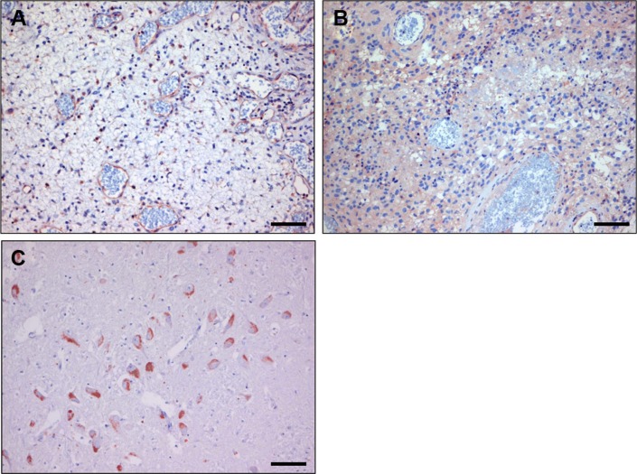
Figure 8. Immunostaining with anti-dynactin-1 -p150Glued of human glioblastoma before (A) and after bevacizumab treatment (B).
Note the similarity with C-C7 immunostainings in Figure 3E and 3F. (Panel C) shows an anti-dynactin staining of normal brain, showing positive neurons, confirming literature data. Bars correspond to 100 μm.

