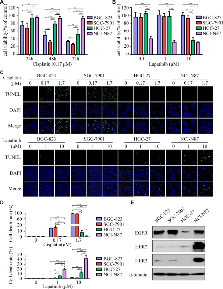
Figure 1. Lapatinib is effective to intrinsic cisplatin-resistant GC cells.
(A) Cell viability was determined by CCK-8 cell-proliferation assay. The gastric cancer (GC) cell lines (BGC-823, SGC-7901, HGC-27, NCI-N87) were exposed to 0.17 μM cisplatin for 24, 48 and 72 h. The percentage of viable cells was shown relative to untreated controls. (B) The cell viabilities of four GC cell lines were measured using CCK-8 assay after 48 h exposure to 0, 0.1, 1 and 10 μM of lapatinib. (C) Cell death was determined by TUNEL assay (× 1000). Top: The four GC cell lines were treated with 0, 0.17, 1.7 μM cisplatin for 24 h. Bottom: The four GC cell lines were treated with 0, 1, 10 μM lapatinib for 24 h. (D) Quantify TUNEL-positive cells of GC cells treated with cisplatin or lapatinib. (E) Western blot showing EGFR, HER2 and HER3 protein expressions in the four GC cell lines. The data presented are means ± SD from three independent experiments. *P < 0.05, **P < 0.01, *** P < 0.001.

