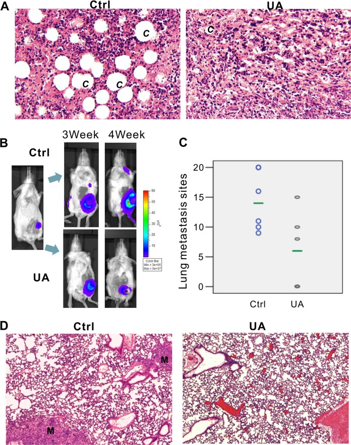
Figure 2. UA inhibits tumor growth and metastasis in vivo.
(A) Hemotoxylin & Eosin staining of 4T1 primary tumors (×200 magnification). 4T1-Luc cells were collected and injected into the mammary fat pad of BALB/c mice (5 × 10⁵ cells/injection; six animals per group). Animals were i.p. injected with 20 mg/kg UA for 4 weeks and sacrificed. Tumor samples were retrieved for H&E staining. C = new capillaries blood vessel in tumors. (B) Xenogen images of representative mouse in control group and UA group. Representative Xenogen images of the mice in 3 weeks and 4 weeks are shown. (C) UA reduced the number of lung metastasis in 4T1 tumor-bearing mice. Picture shown the number of lung metastasis sites on the lung surface for each mouse. The green line shows the average metastasis for each group. (D) Hemotoxylin & Eosin staining of lung tissues for 4T1 tumor-bearing mice and UA treated tumor-bearing mice (×200 magnification). M = metastasis site.

