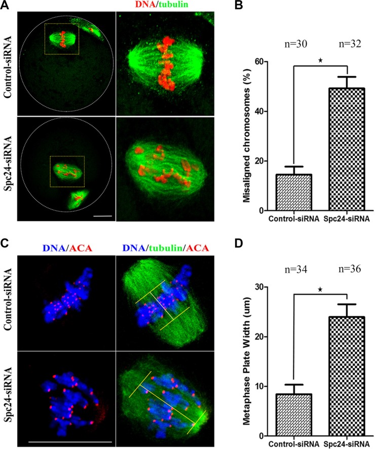
Figure 4. Loss of Spc24 causes misaligned chromosomes in meiotic oocytes.
(A) Abnormal chromosome alignment in MII oocytes after microinjection of Spc24 siRNA. In the control group, most oocytes showed normal chromosome alignment, while in the Spc24-depleted oocytes, most oocytes showed severely misaligned chromosomes. α-tubulin (green); DNA (red). Scale bars: 20 μm. (B) The rates of oocytes with misaligned chromosomes in the siRNA injection and control group. Data are expressed as mean ± SEM of at least 3 independent experiments. *Significantly different (P < 0.05). (C) Oocytes in MI were stained with anti-tubulin, ACA and Hoechst 33342. Scale bars: 20 μm. (D) Metaphase plate width was determined by measuring the axis distance between the two lines at the edges of the DNA. Data are expressed as mean ± SEM of at least 3 independent experiments. *Significantly different (P < 0.05). The total numbers of analyzed oocytes are indicated (n).

