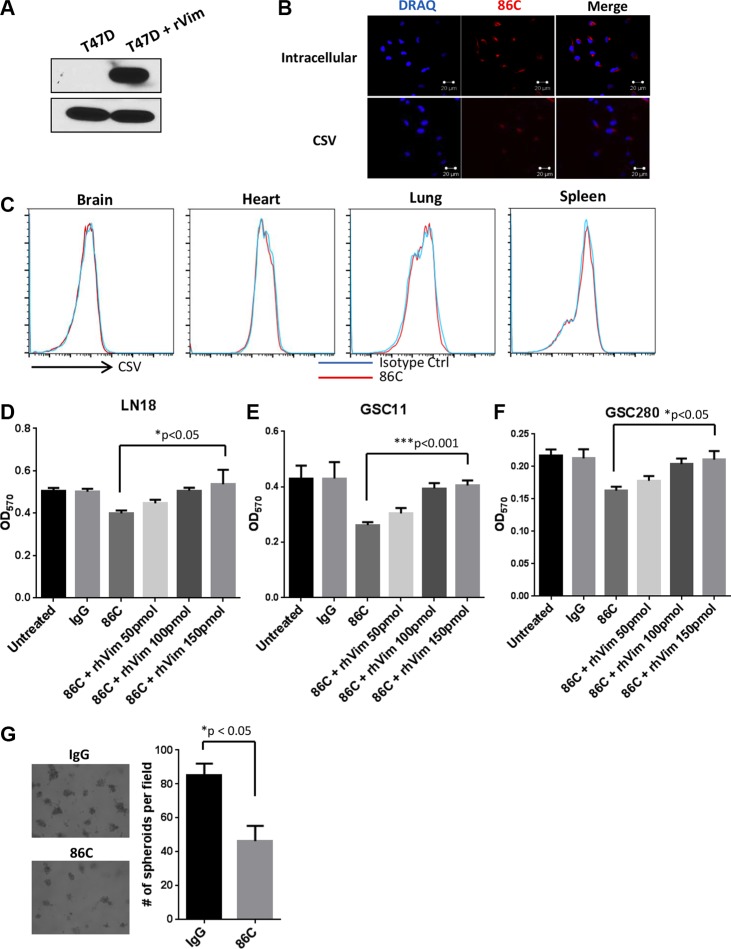
Figure 4. Characterization of the 86C mAb.
(A) T47D cell lysates and T47D cell lysates with 50 ng of rhVim protein were subjected to immunoblotting to detect vimentin using the 86C antibody. GAPDH was used as a loading control. (B) Immunostaining of CSV or cytosolic vimentin (red) expression in LN18 cells. Nuclei (blue) were stained with DRAQ5. (C) Normal mouse cells dissociated from brain, heart, lung, and spleen tissues were stained with mouse IgG or 86C and Alexa Fluor 405–conjugated secondary mouse-IgG. 86C staining was analyzed using flow cytometry. Results are representative of three independent experiments. LN18 (D), GSC11 (E), or GSC280 (F) cells were incubated with various concentrations of rhVim for 1 hour before treatment with 86C to neutralize the impact of 86C on the cells. Cells were collected, and the dead cell population was analyzed using flow cytometry. Data are presented as mean ± standard error (n = 3). *p = 0.02 versus 86C+rhvim 150pmol treatment. Student t test. (G) LN18 cells were suspended on Matrigel in the presence of 86C (10 μg/ml) or IgG. The spheres formed in Matrigel were imaged and counted on day 7.

