
Figure 3. Direct regulation of MTDH by miR-145-5p or miR-145-3p in lung SCC cells.
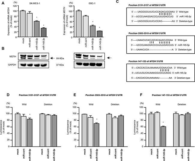
A. MTDH mRNA expression was evaluated by qRT-PCR in SK-MES-1 and EBC-1 cells 72 h after transfection with miR-145-5p or miR-145-3p. GUSB was used as an internal control. *P < 0.0001. B. MTDH protein expression was evaluated by Western blot analyses in SK-MES-1 and EBC-1 72 h after transfection with miR-145-5p or miR-145-3p. GAPDH was used as a loading control. C. miR-145-5p or miR-145-3p binding sites in the 3′-UTR of MTDH mRNA. (D-F) Dual luciferase reporter assays using vectors encoding putative miR-145-5p (positions 3,151-3,157 or 3,503-3,510) or miR-145-3p (147-153) target sites of the MTDH 3′-UTR for both wild-type and deleted regions. Normalized data were calculated as ratios of Renilla/firefly luciferase activities. *P = 0.0054, P < 0.001, and P < 0.001 for D-F., respectively.