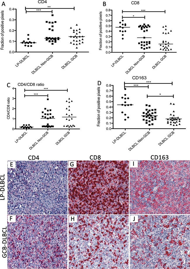
Figure 3. The inflammatory infiltrate in LP-DLBCL shows a low CD4/CD8 ratio and a high content of macrophages.

A. The fraction of CD4-positive pixels was significantly lower in LP-DLBCL compared with Non-GCB and GCB type DLBCL (***p<0.0001, **p=0.0035, Mann-Whitney-test). B. Quantification of the fraction of CD8-positive pixels reveals significantly higher numbers in LP-DLBCL compared with Non-GCB and GCB DLBCL (***p=0.0002, *p=0.0169, Mann-Whitney-test). C. The CD4/CD8 ratio is significantly decreased in the microenvironment of LP-DLBCL compared with Non-GCB and GCB DLBCL (***p≤0.0003, Mann-Whitney-test). D. Quantification of the fraction of CD163-positive pixels reveals significantly higher numbers in LP-DLBCL compared with Non-GCB and GCB DLBCL (***p≤0.0003, *p=0.0109, Mann-Whitney-test). E. Representative example of LP-DLBCL in CD4 immunostaining (200x). F. Representative example of a GCB type DLBCL in CD4 immunostaining (200x). G. Representative example of LP-DLBCL in CD8 immunostaining (200x). H. Representative example of a GCB type DLBCL in CD8 immunostaining (200x). I. Representative example of LP-DLBCL in CD163 immunostaining (200x). J. Representative example of a GCB type DLBCL in CD163 immunostaining (200x).
