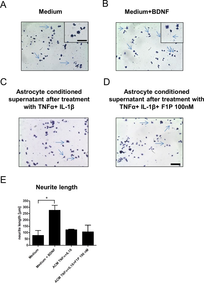
Fig 5. Fingolimod conditioned astrocyte supernatants do not exert growth promoting effects in PC 12 cells.
(A-D) Representative images of PC12 cells in culture after hematoxylin eosin staining. Insets in (A) and (B) show higher manigifcation with representative neurite growth indicated by arrows. Bar indicates 50 µm in D and 20 µm in inset. As compared to (A) medium only as negative control and (B) the addition of BDNF as positive control, the addition of conditioned supernatnats from IL-1β and TNFα inflamed astrocytes with or without F1P treatment at 100 nM (C,D) did not lead to increased neurite lenght. (E) Blinded quantification of neurite lenghts in PC 12 cell culture. ACM, astrocyte conditioned supernatant. Data are given as mean ± SEM, n = 3 per group, 1 out of 2 experiments is shown. * p < 0.05 for medium versus addition of BDNF as positive control, Kruskal Wallis test.

