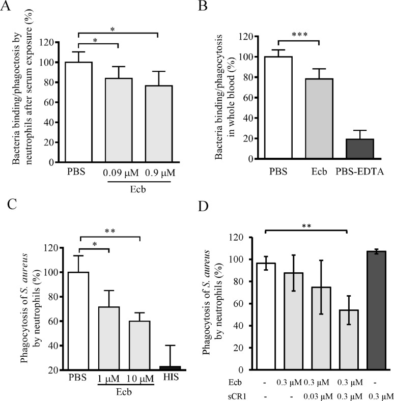
Fig 6. Recognition of opsonized S. aureus and phagocytosis by neutrophils in the presence of Ecb.
A, Fluorescence labeled bacteria were opsonized with C3b by exposure to NHS and mixed with Ecb (0–0.9 μM) prior to analysis of neutrophil-bound or phagocytosed bacteria by flow cytometry. B, The fluorescent labeled bacteria were exposed to hirudin-anticoagulated blood in the presence of Ecb (1.6 μM) prior to flow cytometry. As a control the bacteria were incubated in blood treated with EDTA that blocks complement activity. C, Bacteria labeled with pH rhodoTM were opsonized with C3b by exposure to NHS and incubated with Ecb in the presence of neutrophils. As a control the bacteria were exposed to heat inactivated human serum (HIS, black bar). D, The assay in panel C was done in the presence of 0.3 μM Ecb and increasing concentrations of sCR1. Sample with only sCR1 was used as negative control and sample without any toxin components as positive control for S. aureus phagocytosis by neutrophils. The data are from three, four, and two independent experiments, respectively, with mean SD values. Student’s two-tailed t-test was used to determine the statistical significancies (* p<0.05, ** p<0.01, *** p<0.001).

