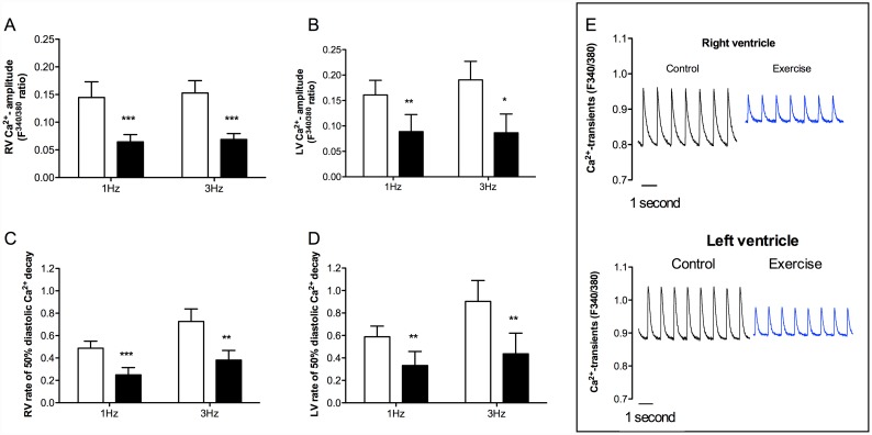
Fig 2. Ca2+ handling in isolated cardiomyocytes from RV and LV of animals subjected to acute exhaustive treadmill exercise (black bars) compared to sedentary controls (white bars).
Stimulation frequencies 1 and 3 Hz, n = 5. (a) Ca2+ transient amplitude in RV; (b) Ca2+ transient amplitude in LV; (c) rate of 50% diastolic Ca2+ decay in RV; (d) rate of 50% diastolic Ca2+ decay in LV; and (e) representative recordings of Ca2+ transients in cardiomyocytes from RV and LV (stimulated at 1 Hz). * p < 0.05, ** p < 0.01, *** p < 0.001.

