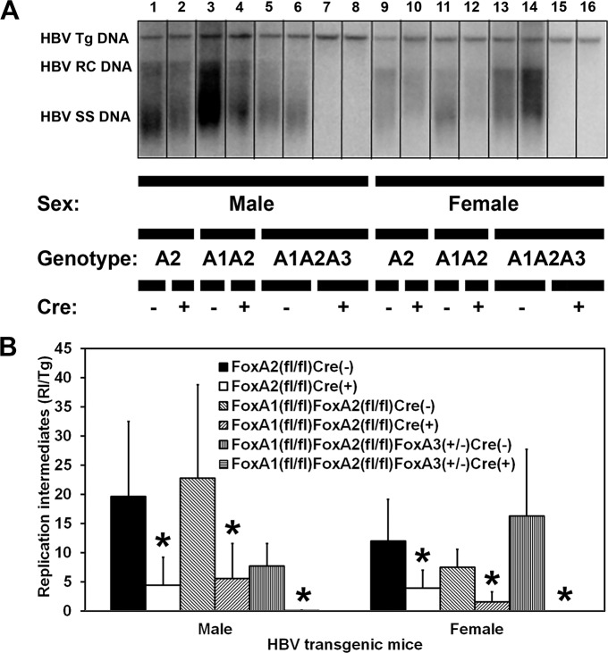
Fig 3. DNA (Southern) filter hybridization analysis of HBV DNA replication intermediates in the livers of adult HBV transgenic mice.
(A) DNA (Southern) filter hybridization analysis of representative mice of each sex and genotype are shown. Noncontiguous lanes from multiple analysis are presented. The probe used was HBVayw genomic DNA. FoxA-expressing (HBVFoxA2fl/flAlbCre(-), HBVFoxA1fl/flFoxA2fl/flAlbCre(-) and HBVFoxA1fl/flFoxA2fl/flFoxA3+/-AlbCre(-)) and FoxA-deleted (HBVFoxA2fl/flAlbCre(+), HBVFoxA1fl/flFoxA2fl/flAlbCre(+) and HBVFoxA1fl/flFoxA2fl/flFoxA3+/-AlbCre(+)) HBV transgenic mice are indicated (Genotype A2, A1A2 and A1A2A3, respectively). The HBV transgene (Tg) was used as an internal control for the quantitation of the HBV replication intermediates. Tg = HBV transgene; RC = HBV relaxed circular replication intermediates; SS = HBV single stranded replication intermediates. (B) Quantitative analysis of the HBV DNA replication intermediate (RI) levels in HBV transgenic mice. The mean DNA replication intermediate levels plus standard deviations are indicated. Average number of mice per group was 6.7±1.8 (Range: 4–9). The levels of replication intermediates which are statistically significantly different between Cre(-) and Cre(+) HBV transgenic mice by a Student’s t-test (p<0.05) are indicated with an asterisk (*).

